menuclose

What is Carotid Artery Stenosis?
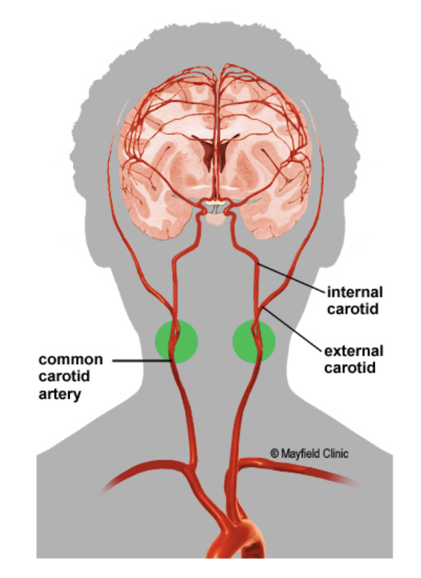
Carotid Artery Stenosis is a disease state that occurs when atherosclerotic plaque builds up within the right and left carotid arteries found in the neck. This obstruction can lead to a reduction of blood flow to the brain, face and neck. Most plaque build-up is typically caused due to higher cholesterol and lipid levels that are deposited within the arterial walls over time.1
Incidence and prevalence of Carotid Artery Stenosis
The incidence of carotid stenosis varies depending on the patients’ age and individual risk factors, however:
- Patients < 50 years of age – approximately 0.5%
- Patients > 70 years of age – approximately 5%
Women and men have similar incidences of carotid stenosis up until 70 years of age when men then appear to have a higher incidence after this age compared to women in the same age group.2
What are the symptoms of Carotid Artery Stenosis?
Carotid artery stenosis may produce little if any symptoms in the earliest stages of the condition and can be undiagnosed for many years until more significant symptoms present. The symptoms are usually a direct result of the fatty deposits within the carotid arteries that reduce the blood flow to the brain, and either temporarily or chronically deprive the brain of blood.
For many patients, the first presentation of this disease can be during a Transient Ischaemic Attack (TIA) or a Stroke. These symptoms may include:
- Severe sudden onset of headache,
- Sudden problems with speech or understanding the speech of others,
- Sudden weakness in any limbs particularly when this occurs on one side of the body,
- Sudden loss of facial muscles or ability to move areas of the face, and
- Sudden loss or change in eyesight or vision particularly when experiencing one-sided issues.
In the case of a TIA, these symptoms may resolve over a short period of time with little to no treatment. However, in the case of a stroke, the symptoms may not resolve without treatment.
Should any of the above symptoms present, this would be considered a medical emergency and patients should immediately go to their nearest hospital or call a local ambulance.4
What are the causes of Carotid Artery Stenosis?
Commonly, carotid artery stenosis is caused by plaque build-up within the arterial wall which may reduce blood flow to the brain, face and neck. This is often associated with Hyperlipidaemia, a condition which leads to atherosclerotic plaque-forming within the blood vessel walls, in turn, reduces the internal dimension of the blood vessel.
Other causes of carotid artery stenosis may include:
- Smoking,
- Ageing,
- Diabetes mellitus, and
- Hypertension.
Occasionally, blood vessels may be damaged if small pieces of plaque crack and rupture. This process can lead to platelets clumping together and forming blood clots around the site of the injury and may reduce blood flow through the carotid artery caused by the newly formed blood clot, leading to symptoms.5
Diagnosis of Carotid Artery Stenosis
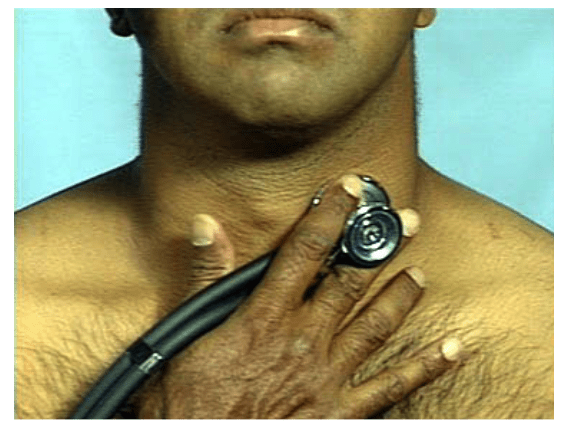
Formal diagnostic tests for carotid artery stenosis are typically performed after a patient has presented or there’s a high suspicion of having either a Transient Ischaemic Attack (TIA) or a Stroke. Occasionally a physician may hear a “bruit” when listening (auscultating) the carotid artery using a stethoscope. This may occur during a physical assessment of a patient by a General Practitioner or another medical specialist.
A “Bruit” may also be referred to as a “murmur” heard when listening to the carotid artery, which can indicate there’s an abnormal flow or obstruction through the arteries.6
Imaging diagnostic options may include:
- Duplex ultrasound scan: uses ultrasound waves to view the arterial walls and measure blood flow through the carotid arteries which identify any potential obstructions of blood flow to the brain
- Computerised tomographic (CT) scan: uses multiple images to construct an image of the arteries and identify any obstructions such as atherosclerotic plaque deposits that may cause symptoms
- Computerised tomographic angiogram (CTA): similar to a CT scan, though a CTA includes a radiopaque dye that is injected and can then be used to highlight arterial blood flow, and spotlighting any obstructions.
- Magnetic Resonance Angiography (MRA): an MRI machine is used to create highly detailed anatomical images which can identify any possible obstructions to blood flow.
- Cerebral angiography: similar to a coronary angiogram, a small catheter is inserted into the artery of interest and contrast dye is injected, recorded and used to identify any narrowing of the carotid artery.7
Classification of Carotid Artery Stenosis
Depending on the level of obstruction that has been identified using specific diagnostic approaches, there are three recognised levels of carotid artery stenosis which can influence treatment options.
- Minor – refers to a narrowing that is < 50% of the width of the carotid artery
- Moderate – refers to any obstruction that causes between 50-69% narrowing of the carotid artery
- Severe – refers to > 70% obstruction of the carotid artery.8
To learn more about the treatment options for Carotid Artery Stenosis, click here.